
Shoulder Dislocation on X-ray: Imaging Findings
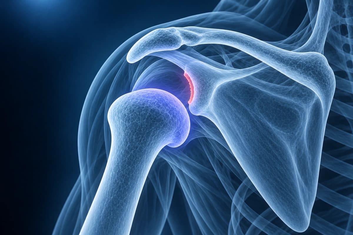
The shoulder (glenohumeral joint) is the most commonly dislocated major joint. Anterior dislocations account for approximately 95% of cases. Imaging after dislocation evaluates for associated injuries including Hill-Sachs lesions (compression fractures of the posterolateral humeral head), Bankart lesions (labral avulsions), glenoid bone loss, and rotator cuff tears.
Common Symptoms
- Severe immediate shoulder pain and inability to move the arm after a traumatic event
- Visible deformity with a flattened deltoid contour and anterior fullness below the clavicle
- The arm held in slight abduction and external rotation in anterior dislocation
- Numbness or tingling in the deltoid patch distribution from axillary nerve traction injury
- Palpable absence of the humeral head in the glenoid fossa
- Apprehension and guarding with any attempted motion of the shoulder
Key Imaging Findings
- Hill-Sachs lesion on the posterolateral humeral head
- Bankart lesion with anteroinferior labral detachment
- Bony Bankart with glenoid rim fracture and bone loss quantification
- Greater tuberosity fracture associated with anterior dislocation
- Rotator cuff tear, especially in patients over 40
- HAGL lesion (humeral avulsion of glenohumeral ligament)
Frequently Asked Questions
What are Hill-Sachs and Bankart lesions and how are they identified on imaging?
A Hill-Sachs lesion is a posterosuperior impaction fracture of the humeral head created when it engages the anterior glenoid rim during anterior dislocation. It appears on axial MRI or CT as a wedge-shaped cortical depression at the posterosuperior humeral head and is best seen on axial T2-FS images or CT with 3D reconstruction. A Bankart lesion is the complementary injury — an anteroinferior labral avulsion, with or without an attached osseous fragment (bony Bankart). MR arthrogram best demonstrates the soft-tissue Bankart lesion as contrast tracking into the labral detachment on axial images. The combination of a large engaging Hill-Sachs lesion and a significant bony Bankart defect increases recurrent instability risk and influences surgical planning.
What is the risk of recurrent dislocation after a first-time shoulder dislocation?
Recurrence risk after a first-time anterior shoulder dislocation is strongly age-dependent. Patients younger than 20 years have recurrence rates approaching 80–90%, while those over 40 years have rates below 15%, partly due to concurrent rotator cuff tears that stabilize the joint. High-demand athletes and military personnel have similarly elevated recurrence risks. MRI findings that predict recurrence include large Hill-Sachs lesions, significant Bankart labral damage, capsular laxity, and anterior glenoid bone loss. Early surgical stabilization with arthroscopic Bankart repair is increasingly recommended for young active individuals after a first-time dislocation to reduce the cumulative damage caused by recurrent episodes.
When is the Latarjet procedure preferred over arthroscopic Bankart repair?
The Latarjet procedure — transfer of the coracoid process with its attached conjoint tendon to the anterior glenoid — is preferred when anterior glenoid bone loss exceeds approximately 20–25% of the glenoid surface, or when an engaging Hill-Sachs lesion creates an off-track bipolar lesion that puts an arthroscopic soft-tissue repair at unacceptably high risk of failure. CT with 3D surface subtraction reconstruction or MRI best quantifies glenoid bone loss. The Latarjet provides a triple stabilizing effect: bony augmentation of the glenoid arc, the sling effect of the conjoint tendon, and the capsulorrhaphy. Recurrence rates with the Latarjet are lower than with soft-tissue repair in high-risk instability cases.
Sources
- Hovelius L, Olofsson A, Sandström B, et al. Nonoperative treatment of primary anterior shoulder dislocation in patients forty years of age and younger. a prospective twenty-five-year follow-up. J Bone Joint Surg Am. 2008;90(5):945-952.
- Robinson CM, Howes J, Murdoch H, et al. Functional outcome and risk of recurrent instability after primary traumatic anterior shoulder dislocation in young patients. J Bone Joint Surg Am. 2006;88(11):2326-2336.
- Burkhart SS, De Beer JF. Traumatic glenohumeral bone defects and their relationship to failure of arthroscopic Bankart repairs: significance of the inverted-pear glenoid and the humeral engaging Hill-Sachs lesion. Arthroscopy. 2000;16(7):677-694.
- Itoi E, Lee SB, Berglund LJ, et al. The effect of a glenoid defect on anteroinferior stability of the shoulder after Bankart repair: a cadaveric study. J Bone Joint Surg Am. 2000;82(1):35-46.
- Di Giacomo G, Itoi E, Burkhart SS. Evolving concept of bipolar bone loss and the Hill-Sachs lesion: from "engaging/non-engaging" lesion to "on-track/off-track" lesion. Arthroscopy. 2014;30(1):90-98.
Related Articles
Your doctor ordered a shoulder MR arthrogram, or the report mentions one? Learn why contrast goes into the joint, what it shows better than MRI and what to ask.
Your shoulder MRI mentions a SLAP tear? Learn what types I to IV mean, from labrum fraying to biceps anchor detachment, and how each type is treated.
After a shoulder dislocation, MRI often shows a Bankart tear and a Hill-Sachs lesion. Learn what they mean, why bone loss matters and the risk of recurrence.
Understand your imaging before your next appointment
MRI, X-ray or CT from a CD, folder or ZIP. A PDF report or photos of the films work too.
Free to open and preview, no account needed. An optional AI report from $10 explains your images in plain language, with questions for your doctor.
Your raw files stay on this device. Only rendered images and minimal context are sent when you request an AI report.
Medical Disclaimer: This page is for informational and educational purposes only. It does not constitute medical advice, diagnosis, or treatment. AI-generated analysis may contain errors. Always consult a qualified healthcare professional for medical decisions. Full Disclaimer