
Rotator Cuff Tear on MRI: Imaging Findings
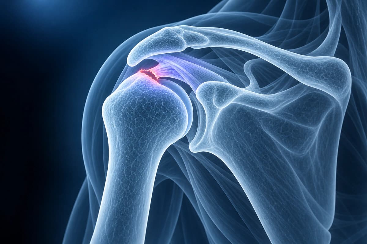
Rotator cuff tears are one of the most common causes of shoulder pain and disability. The rotator cuff comprises four muscles and tendons: supraspinatus (most commonly torn), infraspinatus, teres minor, and subscapularis. MRI is the gold standard for evaluating tear size, location, retraction, muscle quality (fatty infiltration), and associated findings.
Common Symptoms
- Deep aching pain in the shoulder, often worse at night or when lying on the affected side
- Weakness when lifting the arm overhead or rotating the shoulder outward
- Painful arc of motion between 60° and 120° of abduction, classic for supraspinatus tears
- Audible or palpable crepitus with shoulder movement
- Difficulty reaching behind the back or combing hair
- Progressive loss of active range of motion in full-thickness tears
Key Imaging Findings
- Tendon discontinuity or high signal on T2 sequences indicating tear
- Full-thickness tear with tendon gap and possible retraction
- Partial-thickness tear involving articular or bursal surface
- Muscle atrophy and fatty infiltration (Goutallier grading)
- Tendon retraction measurement from the footprint
- Superior migration of the humeral head suggesting massive tear
- Associated biceps tendon pathology or subluxation
Frequently Asked Questions
How is a rotator cuff tear diagnosed on MRI?
MRI is the gold standard for evaluating rotator cuff tears. Coronal-oblique sequences best demonstrate the supraspinatus tendon, while axial T2-FS images reveal the subscapularis and infraspinatus. Full-thickness tears appear as fluid signal completely traversing the tendon on T2-weighted fat-suppressed images. Partial-thickness tears show focal high signal within the tendon substance or at its articular or bursal surface. MR arthrogram with intra-articular gadolinium improves sensitivity for partial-thickness articular-surface tears, as contrast tracks into the tendon defect.
What is the difference between a partial and full-thickness rotator cuff tear?
A partial-thickness tear involves only part of the tendon thickness and does not communicate between the glenohumeral joint and the subacromial bursa. Full-thickness tears extend entirely through the tendon, allowing fluid and, on MR arthrogram, contrast to pass from the joint into the subacromial-subdeltoid bursa. Full-thickness tears are further classified by size: small (<1 cm), medium (1–3 cm), large (3–5 cm), and massive (>5 cm or involving multiple tendons). The supraspinatus is the most commonly torn tendon, typically at its critical zone 1 cm from the footprint.
When is surgery recommended for a rotator cuff tear?
Surgical repair is typically recommended for acute full-thickness tears in active individuals, tears associated with significant weakness or functional loss, and partial tears involving more than 50% of tendon thickness. Younger patients and those with traumatic tears benefit most from early repair, as chronic retraction and fatty infiltration of the supraspinatus and infraspinatus muscles worsen outcomes over time. Arthroscopic techniques allow repair of the torn tendon back to its footprint with suture anchors. Conservative management with physical therapy, corticosteroid injections, and activity modification is first-line for degenerative partial tears in older, lower-demand patients.
Sources
- Goutallier D, Postel JM, Bernageau J, et al. Fatty muscle degeneration in cuff ruptures. Pre- and postoperative evaluation by CT scan. Clin Orthop Relat Res. 1994;(304):78-83.
- Ellman H. Diagnosis and treatment of incomplete rotator cuff tears. Clin Orthop Relat Res. 1990;(254):64-74.
- de Jesus JO, Parker L, Frangos AJ, Nazarian LN. Accuracy of MRI, MR arthrography, and ultrasound in the diagnosis of rotator cuff tears: a meta-analysis. AJR Am J Roentgenol. 2009;192(6):1701-1707.
- American Academy of Orthopaedic Surgeons. Rotator Cuff Tears. OrthoInfo.
Related Articles
Explore whether rotator cuff tears can heal conservatively, factors affecting recovery, and when surgery becomes necessary.
Evidence-based rotator cuff rehabilitation exercises, phases of recovery, and tips for optimal shoulder healing.
Your MRI says partial- or full-thickness rotator cuff tear? Learn what these terms, tear size and retraction mean, and how they shape treatment choices.
Understand your imaging before your next appointment
MRI, X-ray or CT from a CD, folder or ZIP. A PDF report or photos of the films work too.
Free to open and preview, no account needed. An optional AI report from $10 explains your images in plain language, with questions for your doctor.
Your raw files stay on this device. Only rendered images and minimal context are sent when you request an AI report.
Medical Disclaimer: This page is for informational and educational purposes only. It does not constitute medical advice, diagnosis, or treatment. AI-generated analysis may contain errors. Always consult a qualified healthcare professional for medical decisions. Full Disclaimer