
Frozen Shoulder on MRI & X-ray: Imaging Findings
Frozen shoulder, clinically known as adhesive capsulitis, is a condition characterized by progressive stiffness and pain due to inflammation and fibrosis of the glenohumeral joint capsule. It typically evolves through three overlapping stages: the freezing phase (increasing pain and loss of range), the frozen phase (persistent stiffness with gradual pain reduction), and the thawing phase (slow recovery of motion). MRI is valuable for identifying capsular thickening, synovial enhancement, and ruling out other causes of shoulder stiffness. AI can help explain these imaging terms, but a clinician must examine your shoulder to confirm the diagnosis and stage.
Common Symptoms
- Progressive global restriction of both active and passive shoulder range of motion in all planes
- Severe pain in the early freezing phase, disproportionate to the degree of motion loss
- External rotation loss as the earliest and most pronounced deficit, often less than 50% of the contralateral side
- Night pain that disrupts sleep, particularly in the first 3–6 months of the condition
- Gradual spontaneous improvement in the thawing phase over 12–36 months
- Absence of glenohumeral joint space narrowing or other structural pathology on plain radiograph
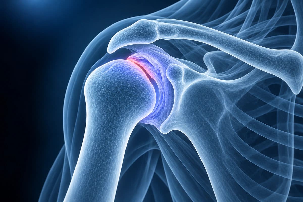
What We Look For on Imaging
- Thickening of the coracohumeral ligament beyond 4 mm on sagittal sequences
- Thickened and enhancing joint capsule at the axillary recess on coronal views
- Capsular enhancement on T1 post-contrast or increased T2 signal in the rotator interval
- Obliteration of the subcoracoid fat triangle indicating rotator interval fibrosis
- Reduced axillary recess volume and inferior capsular scarring
- Exclusion of concurrent rotator cuff tear, labral pathology, or glenohumeral arthritis
Frequently Asked Questions
What does MRI show in adhesive capsulitis?
MRI in adhesive capsulitis (frozen shoulder) demonstrates characteristic thickening of the coracohumeral ligament and rotator interval capsule, reduced axillary pouch volume, and increased T2 signal within the subcoracoid fat triangle on coronal-oblique images. The inferior glenohumeral ligament and axillary recess show capsular thickening and enhancement on contrast-enhanced sequences. MR arthrogram typically reveals reduced joint capacity (normal 20–30 mL, reduced to 5–10 mL in frozen shoulder) with lack of contrast filling the axillary recess and subscapularis bursa. The primary value of MRI in frozen shoulder is to exclude structural pathology such as rotator cuff tears, glenohumeral arthritis, or an occult neoplasm that may mimic the clinical presentation.
What is the natural history of frozen shoulder?
A long-term study found normal or near-normal shoulders in 59% of patients and ongoing symptoms in 41%; most persistent symptoms were mild.
What are the treatment options for frozen shoulder at different stages?
In the freezing phase, intra-articular corticosteroid injections provide the most effective short-term pain relief and accelerate recovery in the early disease course. NSAIDs and analgesics supplement injection therapy. Physical therapy in the acute painful phase must be gentle, as aggressive stretching exacerbates synovial inflammation. In the frozen phase, progressive stretching exercises targeting external rotation, elevation, and internal rotation are the mainstay of rehabilitation. Hydrodilatation — distension of the glenohumeral joint with saline, corticosteroid, and local anesthetic — accelerates recovery in the frozen phase. Manipulation under anesthesia or arthroscopic capsular release is reserved for patients with refractory stiffness beyond 12 months despite conservative measures, releasing the rotator interval and inferior capsule under direct visualization.
Sources
- Reeves B. The natural history of the frozen shoulder syndrome. Scand J Rheumatol. 1975;4(4):193-196.
- Hand C, Clipsham K, Rees JL, Carr AJ. Long-term outcome of frozen shoulder. J Shoulder Elbow Surg. 2008;17(2):231-236.
- Mengiardi B, Pfirrmann CW, Gerber C, et al. Frozen shoulder: MR arthrographic findings. Radiology. 2004;233(2):486-492.
- Challoumas D, Biddle M, McLean M, Millar NL. Comparison of treatments for frozen shoulder: a systematic review and meta-analysis. JAMA Netw Open. 2020;3(12):e2029581.
Understand your imaging before your next appointment
MRI, X-ray or CT from a CD, folder or ZIP. A PDF report or photos of the films work too.
Free to open and preview, no account needed. An optional AI report from $10 explains your images in plain language, with questions for your doctor.
Your raw files stay on this device. Only rendered images and minimal context are sent when you request an AI report.
Medical Disclaimer: This page is for informational and educational purposes only. It does not constitute medical advice, diagnosis, or treatment. AI-generated analysis may contain errors. Always consult a qualified healthcare professional for medical decisions. Full Disclaimer