
Partial vs Full-Thickness Rotator Cuff Tear on MRI
Your MRI says partial- or full-thickness rotator cuff tear? Learn what these terms, tear size and retraction mean, and how they shape treatment choices.
Key Takeaways
- Partial tears affect only part of the tendon thickness; full-thickness tears extend completely through
- Ellman classification grades partial tears by location (articular, bursal, intratendinous) and depth (Grades 1-3)
- Full-thickness tears are sized as small (under 1 cm), medium (1-3 cm), large (3-5 cm), or massive (over 5 cm)
- Patte classification measures tendon retraction, which affects repair feasibility
- Goutallier grading of fatty infiltration (0-4) is critical because it is irreversible and affects surgical outcomes
- Timely treatment prevents progressive fatty infiltration that can make tears irreparable
“Partial-thickness tear” or “full-thickness tear” in your report
A partial-thickness tear goes only part of the way through the tendon, from the joint side (articular-sided) or the bursal side, or sits inside it. A full-thickness tear goes all the way through, so the joint and the bursa above connect. Reports then describe depth or width, retraction and fatty atrophy of the muscle.
How radiologists phrase it
- “Low-grade partial-thickness articular-sided tear of the supraspinatus”
- “High-grade bursal-sided partial-thickness tear involving more than 50% of the tendon”
- “Intrasubstance (interstitial) tear”
- “Full-thickness, partial-width tear of the supraspinatus”
- “Full-thickness tear measuring 1.5 cm with retraction to the humeral head; Goutallier grade 2 fatty infiltration”
Reading the numbers
- Low grade and high grade compare the depth of a partial tear with the tendon's thickness. Ellman's scale grades depth as under 3 mm, 3 to 6 mm, or over 6 mm, which is more than half the tendon.
- “Full-thickness, partial-width” means the tear goes all the way through but only across part of the tendon from front to back.
- Retraction is how far the torn end has pulled back from the bone. Patte staged it from near the insertion, to the humeral head, to the glenoid.
- Fatty atrophy or fatty infiltration (Goutallier grade 0 to 4) describes fat replacing muscle; higher grades are linked with poorer results after repair.
Does it change the plan?
Cuff tears are common in shoulders that do not hurt. One ultrasound study found tears in 13% of people aged 50 to 59 and in 51% of those over 80 without shoulder symptoms, so a report is read alongside pain, strength and function. For full-thickness tears, size, retraction and fatty atrophy are the details surgeons weigh when judging whether a tendon can be repaired.
What to ask at your follow-up
- Is my tear partial or full thickness, and on which side of the tendon?
- How wide is it, and how far has the tendon retracted?
- What grade of fatty change does the muscle show?
- Could my symptoms come from something else on the scan?
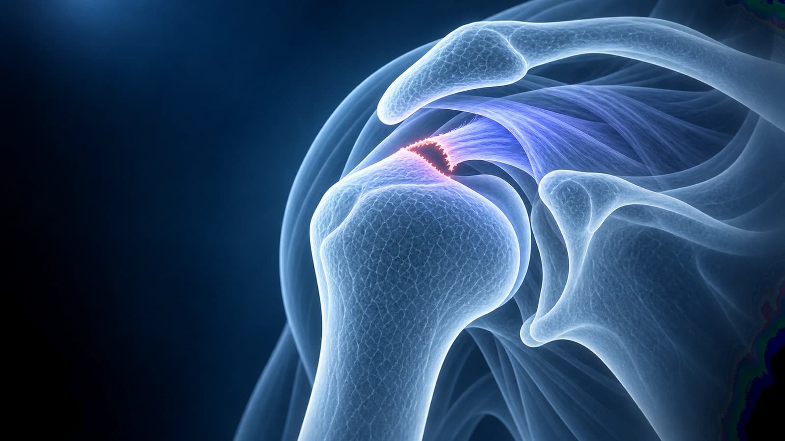
Rotator cuff tears are classified using several grading systems that describe the depth, size, location, and tissue quality of the injury. Understanding these classifications helps you interpret your MRI report, have meaningful conversations with your surgeon, and understand the treatment options available for your specific tear type.
This guide covers the major classification systems used for rotator cuff tears, including the distinction between partial and full-thickness tears, the Ellman classification for partial tears, and the Patte classification for tendon retraction. For guidance on reading your shoulder MRI, see our article on how to read shoulder MRI.
Partial vs Full-Thickness Tears
The most fundamental distinction in rotator cuff tears is between partial-thickness and full-thickness injuries. A partial-thickness tear involves only part of the tendon depth — the defect does not extend completely through the tendon from one surface to the other. A full-thickness tear means the defect extends all the way through, creating a communication between the joint space below and the subacromial bursa above. On MRI, partial tears show focal bright signal on T2 images that affects only part of the tendon thickness, while full-thickness tears show bright signal extending completely through the tendon.
Partial-Thickness Tear Classification (Ellman)
The Ellman classification grades partial tears by location (Type A: articular-side, accounting for 60% of partial tears; Type B: bursal-side; Type C: intratendinous) and depth:
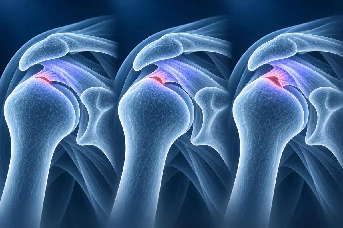
- Grade 1: less than 3 mm deep (less than 25% of tendon thickness)
- Grade 2: 3-6 mm deep (approximately 25-50% of tendon thickness)
- Grade 3: greater than 6 mm deep (more than 50% of tendon thickness)
Full-Thickness Tear Size Classification
Full-thickness tears are classified by their size measured in the anteroposterior dimension (front to back):
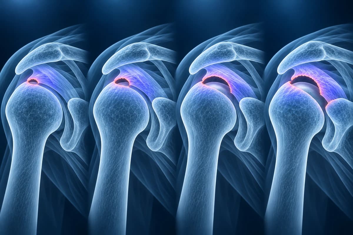
- Small: less than 1 cm — often responds well to conservative treatment
- Medium: 1-3 cm — may require surgery depending on symptoms and demands
- Large: 3-5 cm — typically requires surgical repair for active patients
- Massive: greater than 5 cm or involving two or more tendons — challenging to repair, may be irreparable
Patte Classification (Tendon Retraction)
The Patte classification describes the degree of tendon retraction in full-thickness tears — how far the torn tendon end has pulled away from its insertion on the humerus. This is assessed on coronal oblique MRI images:
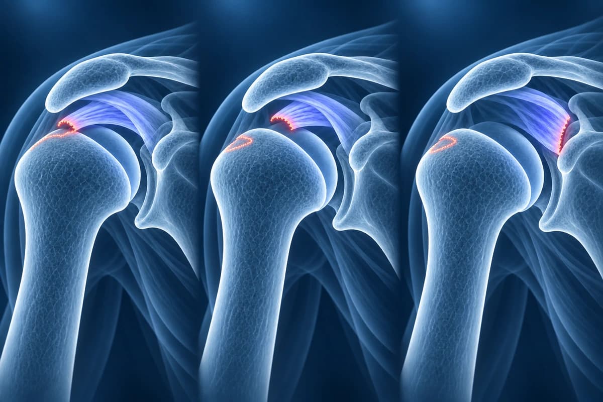
- Stage 1: tendon stump near insertion (proximal to bony insertion) — good repair potential
- Stage 2: tendon stump retracted to the level of the humeral head — moderate repair difficulty
- Stage 3: tendon stump retracted to the level of the glenoid — difficult or impossible to repair primarily
Goutallier Classification (Fatty Infiltration)
When a rotator cuff tendon is torn, the muscle may undergo fatty infiltration — graded 0 (normal) to 4 (more fat than muscle) on sagittal T1 MRI. Grades 3-4 are largely irreversible and associated with poor surgical outcomes, potentially making tears irreparable. Early evaluation is important to prevent progressive infiltration.
Frequently Asked Questions
Can a partial rotator cuff tear become a full tear?
Yes. Partial tears can progress to full-thickness tears over time, particularly high-grade partial tears (Ellman Grade 3, greater than 50% thickness). Studies suggest approximately 25-50% of partial tears will progress over a 2-year period, especially in patients who continue high-demand overhead activities. Regular monitoring with ultrasound or MRI can track tear progression.
What makes a rotator cuff tear irreparable?
A tear is considered irreparable when the tendon has retracted significantly (Patte Stage 3), the muscle has undergone advanced fatty infiltration (Goutallier Grade 3-4), and the tissue quality is too poor to hold sutures. In these cases, alternative procedures such as superior capsular reconstruction, tendon transfer, or reverse total shoulder replacement may be considered.
Do all rotator cuff tears need surgery?
No. Studies show 60-80% of patients achieve satisfactory outcomes with physical therapy alone. Surgery is recommended for large tears in active patients and tears failing conservative treatment. See our shoulder rehab guide.
Sources
- Ellman H. Diagnosis and treatment of incomplete rotator cuff tears. Clin Orthop Relat Res. 1990;(254):64-74.
- Patte D. Classification of rotator cuff lesions. Clin Orthop Relat Res. 1990;(254):81-86.
- Goutallier D, Postel JM, Bernageau J, et al. Fatty muscle degeneration in cuff ruptures. Pre- and postoperative evaluation by CT scan. Clin Orthop Relat Res. 1994;(304):78-83.
- Fuchs B, Weishaupt D, Zanetti M, et al. Fatty degeneration of the muscles of the rotator cuff: assessment by computed tomography versus magnetic resonance imaging. J Shoulder Elbow Surg. 1999;8(6):599-605.
- DeOrio JK, Cofield RH. Results of a second attempt at surgical repair of a failed initial rotator-cuff repair. J Bone Joint Surg Am. 1984;66(4):563-567.
- Tempelhof S, Rupp S, Seil R. Age-related prevalence of rotator cuff tears in asymptomatic shoulders. J Shoulder Elbow Surg. 1999;8(4):296-299.
- Morag Y, Jacobson JA, Miller B, et al. MR imaging of rotator cuff injury: what the clinician needs to know. Radiographics. 2006;26(4):1045-1065.
Related Articles
Explore whether rotator cuff tears can heal conservatively, factors affecting recovery, and when surgery becomes necessary.
A patient's guide to the shoulder MRI report: rotator cuff, labrum, biceps and bursa terms in plain words, and what common findings usually mean.
Related Conditions
Understand your imaging before your next appointment
MRI, X-ray or CT from a CD, folder or ZIP. A PDF report or photos of the films work too.
Free to open and preview, no account needed. An optional AI report from $10 explains your images in plain language, with questions for your doctor.
Your raw files stay on this device. Only rendered images and minimal context are sent when you request an AI report.
Medical Disclaimer: This page is for informational and educational purposes only. It does not constitute medical advice, diagnosis, or treatment. AI-generated analysis may contain errors. Always consult a qualified healthcare professional for medical decisions. Full Disclaimer