
Shoulder MR Arthrogram Explained: What the Report Means
Your doctor ordered a shoulder MR arthrogram, or the report mentions one? Learn why contrast goes into the joint, what it shows better than MRI and what to ask.
What a shoulder MR arthrogram is
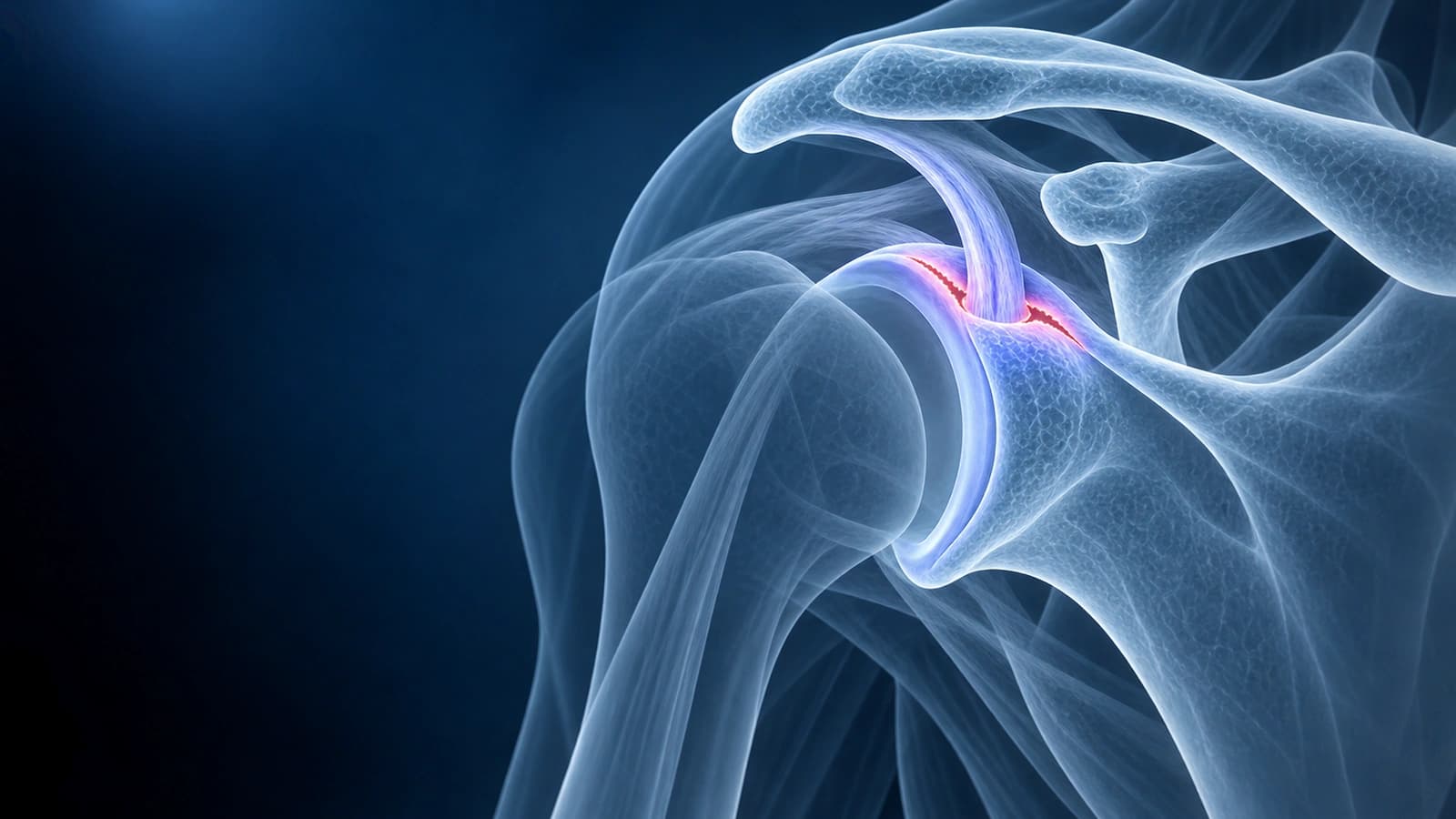
A shoulder MR arthrogram is an MRI taken after diluted contrast, usually gadolinium, or saline is injected into the shoulder joint under imaging guidance. The fluid distends the joint and outlines the labrum, the capsule and the underside of the rotator cuff, so tears that a standard MRI can miss are easier to see.
Phrases from an arthrogram report
- “Intra-articular injection of dilute gadolinium under fluoroscopic guidance”
- “Contrast extends into the superior labrum, consistent with a SLAP tear”
- “No contrast in the subacromial-subdeltoid bursa to suggest a full-thickness tear”
- “Indirect MR arthrogram”, meaning contrast was given into a vein instead of into the joint
What it shows better than a standard MRI
- Labral tears: in a meta-analysis of 60 studies, MR arthrography found labral lesions with 88% sensitivity, compared with 76% for standard MRI.
- Partial versus full-thickness cuff tears: contrast that passes from the joint into the bursa above the cuff shows that a tear goes all the way through.
- Normal variants: a sublabral recess, which can mimic a SLAP tear, is shown more clearly than on MRI without contrast.
Safety, and why it is ordered
Arthrography is considered very safe. In a survey of experienced radiologists covering about 13,300 MR arthrograms, six reactions were reported, all minor, such as pain, fainting reactions and headaches. It is usually chosen when a labral tear or shoulder instability is suspected, or when a standard MRI left a question open.
Questions before or after the arthrogram
- What question should the arthrogram answer that my MRI did not?
- Will the injection be guided by fluoroscopy or ultrasound, and how long does it take?
- Did contrast enter the labrum or the bursa, and what does that mean for me?
- Should the team know about any allergies or earlier reactions to contrast?
Sources
- Steinbach LS, Palmer WE, Schweitzer ME. Special focus session. MR arthrography. Radiographics. 2002;22(5):1223-1246.
- Smith TO, Drew BT, Toms AP. A meta-analysis of the diagnostic test accuracy of MRA and MRI for the detection of glenoid labral injury. Arch Orthop Trauma Surg. 2012;132(7):905-919.
- Smith DK, Chopp TM, Aufdemorte TB, et al. Sublabral recess of the superior glenoid labrum: study of cadavers with conventional nonenhanced MR imaging, MR arthrography, anatomic dissection, and limited histologic examination. Radiology. 1996;201(1):251-256.
- Hugo PC 3rd, Newberg AH, Newman JS, Wetzner SM. Complications of arthrography. Semin Musculoskelet Radiol. 1998;2(4):345-348.
- Expert Panel on Musculoskeletal Imaging; Amini B, Beckmann NM, et al. ACR Appropriateness Criteria Shoulder Pain-Traumatic. J Am Coll Radiol. 2018;15(5S):S171-S188.
Related Articles
Your shoulder MRI mentions a SLAP tear? Learn what types I to IV mean, from labrum fraying to biceps anchor detachment, and how each type is treated.
After a shoulder dislocation, MRI often shows a Bankart tear and a Hill-Sachs lesion. Learn what they mean, why bone loss matters and the risk of recurrence.
Related Conditions
Understand your imaging before your next appointment
MRI, X-ray or CT from a CD, folder or ZIP. A PDF report or photos of the films work too.
Free to open and preview, no account needed. An optional AI report from $10 explains your images in plain language, with questions for your doctor.
Your raw files stay on this device. Only rendered images and minimal context are sent when you request an AI report.
Medical Disclaimer: This page is for informational and educational purposes only. It does not constitute medical advice, diagnosis, or treatment. AI-generated analysis may contain errors. Always consult a qualified healthcare professional for medical decisions. Full Disclaimer