
Shoulder Arthritis on X-ray: Imaging Findings
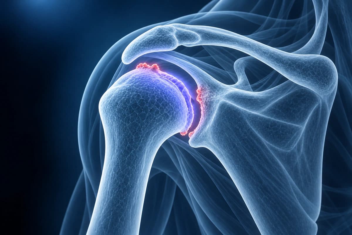
Shoulder arthritis can affect both the glenohumeral joint and the acromioclavicular (AC) joint. Glenohumeral osteoarthritis is characterized by progressive cartilage loss, osteophyte formation, and glenoid wear. AC joint arthritis is extremely common and often found incidentally. Imaging plays a critical role in evaluating joint space, cartilage status, glenoid morphology (Walch classification), and associated rotator cuff status. AI can help explain arthritis-related imaging terms, but a shoulder specialist must assess treatment options, including whether replacement surgery is appropriate.
Common Symptoms
- Deep, aching glenohumeral pain that worsens progressively over months to years
- Stiffness and loss of range of motion, particularly internal rotation and cross-body adduction
- Painful grinding or crepitus with shoulder movement
- Night pain and difficulty sleeping on the affected side
- Weakness secondary to disuse atrophy of the surrounding musculature
- Swelling and warmth of the shoulder joint in inflammatory arthritis
Key Imaging Findings
- Glenohumeral joint space narrowing on X-ray
- Inferior humeral head osteophytes (goat's beard osteophyte)
- Posterior glenoid wear with biconcavity (Walch classification)
- Subchondral cyst formation and sclerosis
- AC joint narrowing, osteophytes, and inferior spur formation
- Rotator cuff status assessment for arthroplasty planning
Frequently Asked Questions
What are the different types of shoulder arthritis and how do they differ on imaging?
Primary glenohumeral osteoarthritis shows characteristic findings on X-ray and MRI: joint space narrowing, subchondral sclerosis, osteophytes at the inferior humeral head and glenoid, and posterior glenoid erosion with posterior humeral head subluxation (the Walch classification). Rheumatoid arthritis presents with symmetric joint space loss, periarticular erosions, and osteopenia without large osteophytes. Rotator cuff arthropathy is characterized by superior migration of the humeral head, acetabularization of the acromion, and femoralization of the humeral head due to chronic massive rotator cuff tear. Post-traumatic arthritis can show irregular joint surfaces, hardware from prior surgery, and malunion deformity. MRI additionally demonstrates cartilage thickness and rotator cuff integrity, critical for surgical planning.
How does MRI help plan shoulder replacement surgery?
Preoperative MRI provides information not available on plain radiographs that directly impacts implant selection and surgical technique. Assessment of rotator cuff integrity on coronal-oblique and axial T2-FS sequences determines whether an anatomic total shoulder arthroplasty (requiring intact rotator cuff) or a reverse total shoulder arthroplasty (suitable for cuff-deficient shoulders) is appropriate. Glenoid morphology and version are evaluated on axial images to guide glenoid component positioning and the need for bone grafting. The degree of posterior glenoid erosion and humeral head subluxation on axial sequences helps quantify deformity correction required. Muscle quality of the rotator cuff, deltoid, and periscapular muscles informs rehabilitation prognosis.
What non-surgical treatments are available for shoulder arthritis?
Conservative management of glenohumeral arthritis includes NSAIDs for analgesic and anti-inflammatory effect, activity modification to avoid high-impact loading, and physical therapy to maintain range of motion and strengthen periarticular musculature. Intra-articular corticosteroid injections provide temporary relief lasting weeks to months and may be repeated two to three times per year. Viscosupplementation with hyaluronic acid is used off-label by some practitioners, though evidence for glenohumeral arthritis is less robust than for the knee. Platelet-rich plasma injections are under investigation. Total shoulder or reverse shoulder arthroplasty is the definitive treatment for end-stage arthritis refractory to non-surgical measures, with excellent pain relief and functional outcomes.
Sources
- Walch G, Badet R, Boulahia A, Khoury A. Morphologic study of the glenoid in primary glenohumeral osteoarthritis. J Arthroplasty. 1999;14(6):756-760.
- Neer CS 2nd, Craig EV, Fukuda H. Cuff-tear arthropathy. J Bone Joint Surg Am. 1983;65(9):1232-1244.
- Hamada K, Fukuda H, Mikasa M, Kobayashi Y. Roentgenographic findings in massive rotator cuff tears. A long-term observation. Clin Orthop Relat Res. 1990;(254):92-96.
- American Academy of Orthopaedic Surgeons. Arthritis of the Shoulder. OrthoInfo.
Understand your imaging before your next appointment
MRI, X-ray or CT from a CD, folder or ZIP. A PDF report or photos of the films work too.
Free to open and preview, no account needed. An optional AI report from $10 explains your images in plain language, with questions for your doctor.
Your raw files stay on this device. Only rendered images and minimal context are sent when you request an AI report.
Medical Disclaimer: This page is for informational and educational purposes only. It does not constitute medical advice, diagnosis, or treatment. AI-generated analysis may contain errors. Always consult a qualified healthcare professional for medical decisions. Full Disclaimer