
Biceps Tendon Injury on MRI: Imaging Findings
The long head of the biceps tendon (LHBT) originates from the superior glenoid labrum and supraglenoid tubercle, courses through the glenohumeral joint, and exits through the bicipital groove. LHBT pathology includes tendinopathy, partial and complete tears, subluxation, and dislocation from the groove. These conditions often coexist with rotator cuff tears and labral injuries.
Common Symptoms
- Anterior shoulder pain localized to the bicipital groove on palpation
- Pain with resisted forearm supination and elbow flexion (Speed's and Yergason's tests)
- A visible "Popeye" deformity with distal migration of the biceps muscle belly after proximal rupture
- Sudden pop followed by pain and bruising in the arm at the time of tendon rupture
- Weakness with elbow flexion and supination, more pronounced with complete rupture
- Deep aching in the anterior shoulder with overhead activities in tendinopathy
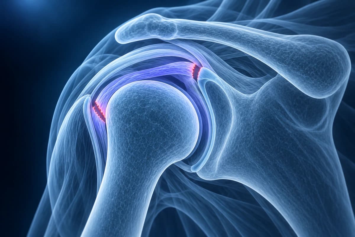
Key Imaging Findings
- Increased signal within the LHBT indicating tendinopathy or tear
- Tendon subluxation or dislocation from the bicipital groove
- Complete tendon rupture with empty groove sign
- Biceps pulley injury with medial subluxation
- Fluid in the biceps tendon sheath (tenosynovitis)
- Associated subscapularis tear allowing biceps medial dislocation
Frequently Asked Questions
How is long head of biceps tendon pathology assessed on MRI?
The long head of the biceps tendon (LHBT) is best evaluated on axial MRI sequences as it courses through the bicipital groove. Normal tendon appears as a uniformly dark round structure on T2-weighted images. Tendinopathy shows intratendinous T2 hyperintensity and tendon thickening. Tenosynovitis is identified by fluid surrounding the tendon within the bicipital groove sheath, distinct from a small amount of physiologic fluid that communicates with the glenohumeral joint. Complete rupture manifests as absence of the tendon within the groove and is confirmed by identifying the retracted tendon end. Axial T2-FS images at the level of the groove and coronal-oblique sequences at the superior labrum biceps anchor are complementary for full assessment.
What is the relationship between biceps tendon pathology and SLAP tears?
The long head of the biceps tendon originates from the supraglenoid tubercle and the superior labrum, making SLAP tears and biceps anchor pathology intimately related. Traction on the biceps tendon during deceleration of the overhead throw transmits load to the superior labrum, producing type II SLAP tears at the biceps anchor — the most common type in overhead athletes. MR arthrogram with intra-articular gadolinium is the preferred study to assess both the biceps anchor and the adjacent superior labrum on coronal-oblique and sagittal sequences. Intrasubstance signal change in the proximal biceps tendon on axial images often accompanies superior labral tears. Surgical decision-making between SLAP repair and biceps tenodesis depends on patient age, activity demands, and the degree of intratendinous biceps degeneration.
When is biceps tenodesis preferred over SLAP repair?
Biceps tenodesis — detaching the LHBT from its superior labral origin and reattaching it to the proximal humerus — is preferred over SLAP repair in patients over 35 years of age, those with significant intratendinous biceps degeneration, workers performing repetitive forearm supination, and patients in whom the SLAP tear is associated with concurrent bicipital groove pathology. In older patients, SLAP repair carries higher failure rates and longer recovery. Tenodesis reliably eliminates anterior shoulder pain arising from the biceps-labral complex while preserving biceps function. Subpectoral or arthroscopic suprapectoral tenodesis locations each have advantages; the subpectoral approach removes the entire pathologic intertubercular segment of the tendon. SLAP repair remains preferred for young overhead athletes with isolated type II SLAP tears.
Sources
- Nho SJ, Strauss EJ, Lenart BA, et al. Long head of the biceps tendinopathy: diagnosis and management. J Am Acad Orthop Surg. 2010;18(11):645-656.
- Elser F, Braun S, Dewing CB, et al. Anatomy, function, injuries, and treatment of the long head of the biceps brachii tendon. Arthroscopy. 2011;27(4):581-592.
- Boileau P, Parratte S, Chuinard C, et al. Arthroscopic treatment of isolated type II SLAP lesions: biceps tenodesis as an alternative to reinsertion. Am J Sports Med. 2009;37(5):929-936.
- Provencher MT, McCormick F, Dewing C, et al. A prospective analysis of 179 type 2 superior labrum anterior and posterior repairs: outcomes and factors associated with success and failure. Am J Sports Med. 2013;41(4):880-886.
Related Articles
Learn when 3T shoulder MRI can help for labral tears, small rotator cuff tears, instability, and cartilage detail, and when 1.5T MRI or ultrasound is enough.
A patient's guide to the shoulder MRI report: rotator cuff, labrum, biceps and bursa terms in plain words, and what common findings usually mean.
Understand your imaging before your next appointment
MRI, X-ray or CT from a CD, folder or ZIP. A PDF report or photos of the films work too.
Free to open and preview, no account needed. An optional AI report from $10 explains your images in plain language, with questions for your doctor.
Your raw files stay on this device. Only rendered images and minimal context are sent when you request an AI report.
Medical Disclaimer: This page is for informational and educational purposes only. It does not constitute medical advice, diagnosis, or treatment. AI-generated analysis may contain errors. Always consult a qualified healthcare professional for medical decisions. Full Disclaimer