
AC Joint Separation on MRI & X-ray: Imaging Findings
Acromioclavicular (AC) joint separation results from disruption of the ligaments stabilizing the junction between the clavicle and the acromion. It is classified using the Rockwood system from Type I (sprain of the AC ligaments) through Type VI (inferior dislocation of the clavicle). Types I-III are most common and result from falls onto the shoulder or direct impact. Imaging with MRI and radiographs is essential for grading severity, assessing ligament integrity, and identifying associated injuries.
Common Symptoms
- Acute pain and tenderness directly over the acromioclavicular joint after a direct blow or fall
- Visible step deformity at the AC joint with superior prominence of the distal clavicle in higher-grade injuries
- Pain with cross-body adduction (horizontal adduction test) bringing the arm across the chest
- Weakness and discomfort with overhead pressing and lifting activities
- A palpable or visible "piano key" sign with downward pressure on the distal clavicle in type IV–VI injuries
- Bruising and swelling over the superior shoulder in acute moderate-to-severe separations
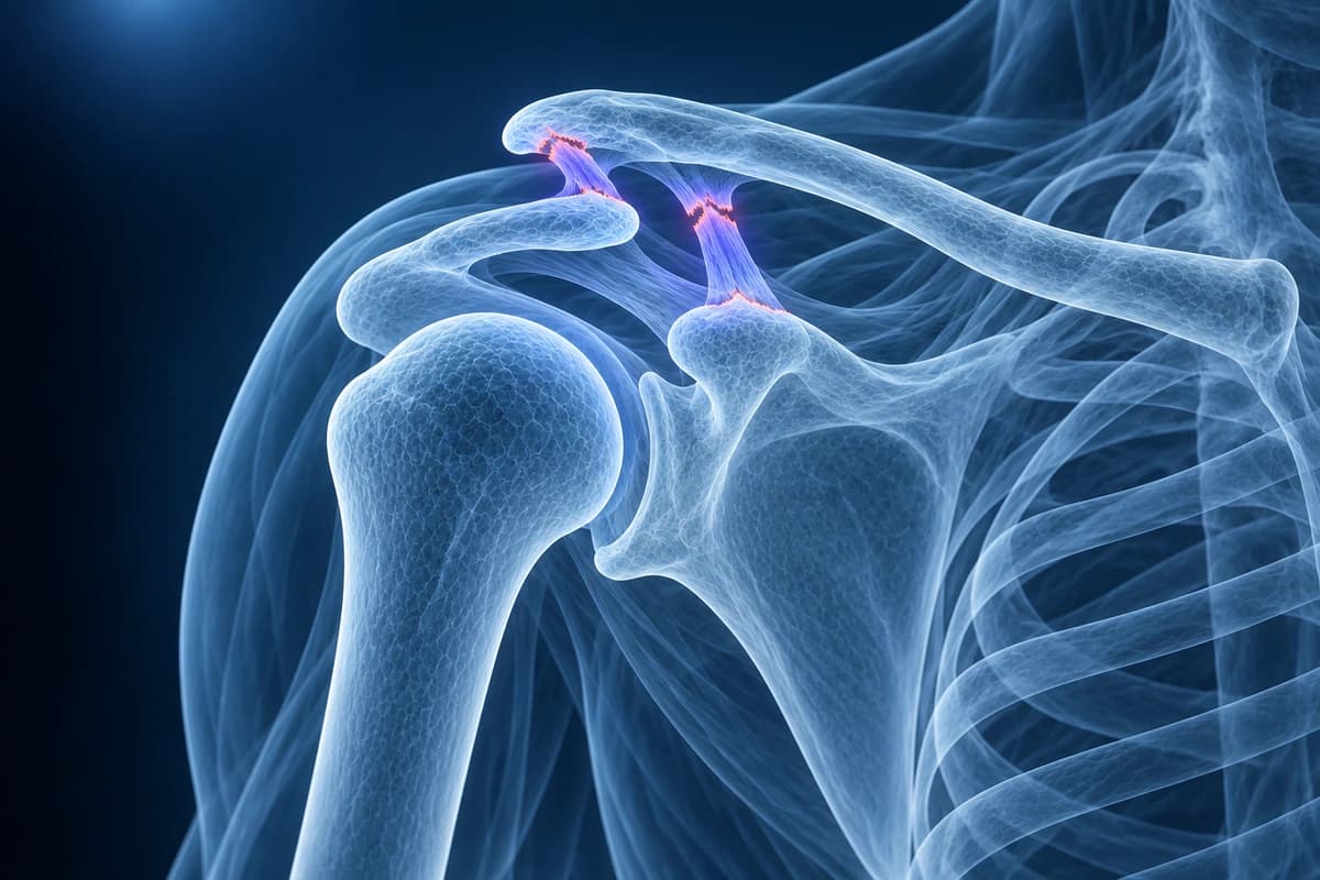
What We Look For on Imaging
- Widening of the AC joint space beyond 7 mm indicating ligament disruption
- Increased coracoclavicular distance compared to the contralateral side
- High signal or discontinuity of AC and coracoclavicular ligaments on MRI
- Superior displacement of the distal clavicle relative to the acromion
- Periarticular edema and joint effusion on fluid-sensitive sequences
- Associated injuries to the deltotrapezial fascia or rotator cuff
Frequently Asked Questions
How is AC joint separation classified and what imaging determines severity?
AC separations are classified by ligament injury and clavicle displacement. Type I is an AC ligament sprain; type II adds AC ligament rupture and CC ligament injury; type III disrupts both. Types IV, V, and VI describe posterior, marked superior, and inferior displacement. Examination and X-rays assess alignment; type I can look normal on X-rays.
Which AC joint separations require surgery?
Types I and II AC separations are treated conservatively with sling immobilization, ice, analgesics, and progressive physical therapy, with excellent functional outcomes. Type III injuries are the most controversial — the majority of patients treated nonoperatively return to full function, while surgery is reserved for high-demand overhead athletes, manual laborers, and those with persistent symptomatic instability after 3 months of conservative management. Types IV, V, and VI — with posterior, markedly superior (>100% CC distance), or inferior-subcoracoid displacement respectively — are treated with surgical stabilization. Surgical techniques include hook plate fixation, coracoclavicular ligament reconstruction with synthetic or autograft tissue, and AC joint stabilization with suture button devices. Concurrent rotator cuff or labral tears identified on MRI are addressed arthroscopically at the same time.
What is the long-term prognosis after AC joint separation?
The majority of patients with type I and II AC separations recover fully within 6–12 weeks with conservative management. Type III injuries treated nonoperatively achieve satisfactory outcomes in 80–90% of patients, although persistent AC joint prominence and mild functional deficits are common. Chronic AC joint pain from post-traumatic osteoarthritis of the AC joint can develop years after any grade of separation, presenting as pain with cross-body adduction and overhead activities. Distal clavicle resection, performed arthroscopically, reliably alleviates chronic AC joint arthritis pain. High-grade injuries treated with surgical stabilization have good return-to-sport rates, though hardware complications, loss of reduction, and coracoclavicular ossification are recognized risks.
Sources
- Simovitch R, Sanders B, Ozbaydar M, et al. Acromioclavicular joint injuries: diagnosis and management. J Am Acad Orthop Surg. 2009;17(4):207-219.
- Petersson CJ, Redlund-Johnell I. Radiographic joint space in normal acromioclavicular joints. Acta Orthop Scand. 1983;54(3):431-433.
- Phillips AM, Smart C, Groom AF. Acromioclavicular dislocation. Conservative or surgical therapy. Clin Orthop Relat Res. 1998;(353):10-17.
- American Academy of Orthopaedic Surgeons. Shoulder Separation. OrthoInfo.
Understand your imaging before your next appointment
MRI, X-ray or CT from a CD, folder or ZIP. A PDF report or photos of the films work too.
Free to open and preview, no account needed. An optional AI report from $10 explains your images in plain language, with questions for your doctor.
Your raw files stay on this device. Only rendered images and minimal context are sent when you request an AI report.
Medical Disclaimer: This page is for informational and educational purposes only. It does not constitute medical advice, diagnosis, or treatment. AI-generated analysis may contain errors. Always consult a qualified healthcare professional for medical decisions. Full Disclaimer