
Thoracic Outlet Syndrome on MRI & X-ray: Imaging Findings
Thoracic outlet syndrome (TOS) refers to compression of the neurovascular bundle — the brachial plexus, subclavian artery, or subclavian vein — as it passes through the thoracic outlet between the scalene muscles, first rib, and clavicle. Causes include cervical ribs, scalene muscle hypertrophy, fibrous bands, and post-traumatic changes. It is classified into neurogenic (most common, 95%), venous, and arterial subtypes. MRI is valuable for identifying anatomic variants, muscular abnormalities, and vascular compression.
Common Symptoms
- Aching pain, heaviness, and fatigue in the shoulder and arm that worsens with overhead activities
- Numbness, tingling, or paresthesia in the ulnar forearm and fourth and fifth fingers (C8-T1 distribution)
- Weakness and wasting of intrinsic hand muscles in chronic neurogenic thoracic outlet syndrome
- Arm swelling, discoloration, and distended superficial veins in venous (Paget-Schroetter) thoracic outlet syndrome
- Ischemic symptoms including pallor, coldness, and digital ischemia in arterial thoracic outlet syndrome
- Symptom reproduction with provocative positioning: arm elevation, the Adson maneuver, or EAST test
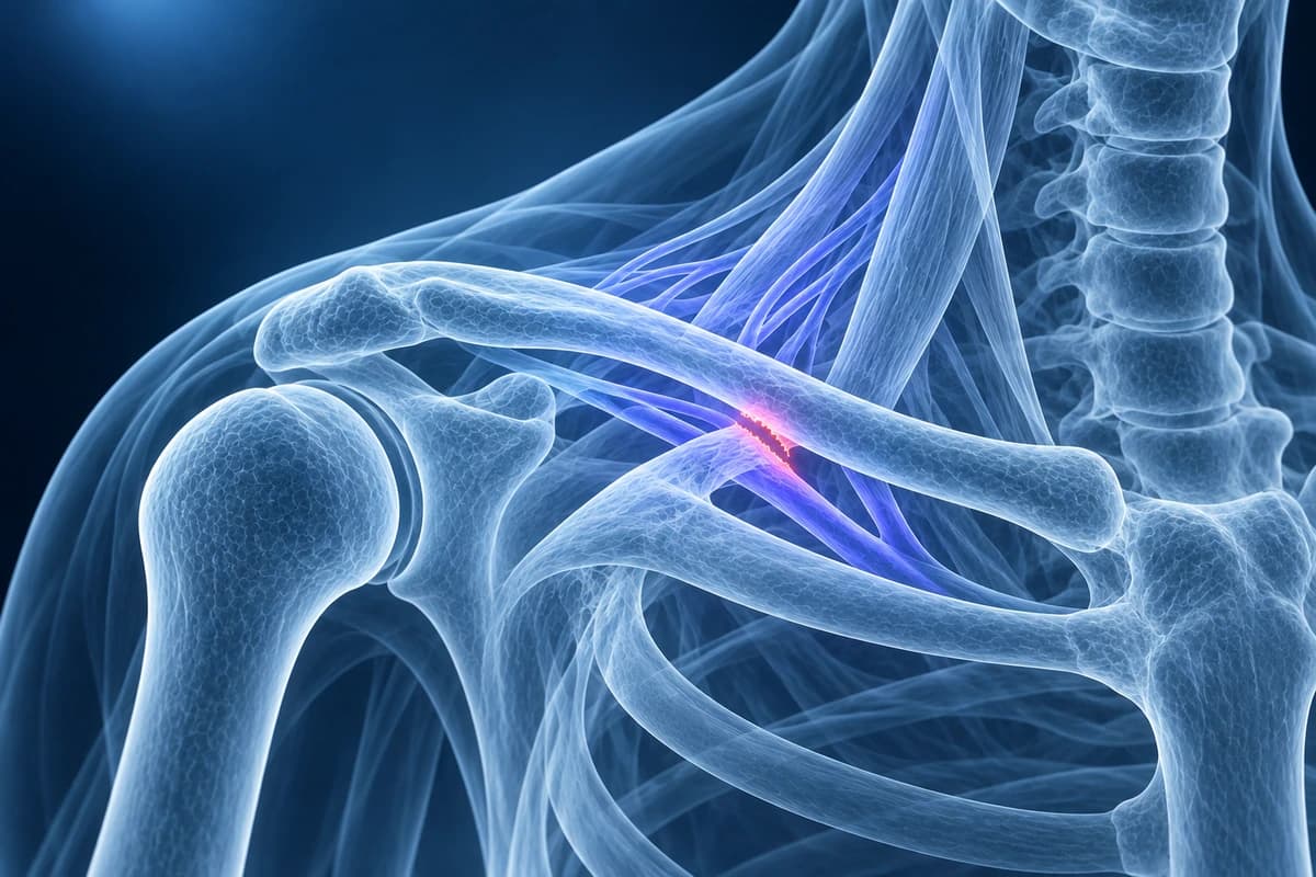
What We Look For on Imaging
- Presence of a cervical rib or elongated C7 transverse process
- Scalene muscle hypertrophy or asymmetry narrowing the interscalene triangle
- Fibrous bands or anomalous soft tissue within the thoracic outlet
- Compression or displacement of the subclavian artery or vein
- Brachial plexus edema or signal abnormality on MRI neurography sequences
- Narrowing of the costoclavicular space between the clavicle and first rib
Frequently Asked Questions
What imaging studies are used to evaluate thoracic outlet syndrome?
Plain radiographs are the first-line study to identify cervical ribs or an elongated C7 transverse process compressing the brachial plexus or subclavian vessels. CT angiography or MR angiography with the arms in neutral and provocative elevated positions delineates arterial and venous compression dynamically. MRI of the brachial plexus using dedicated neurography sequences (coronal STIR and 3D FIESTA or SPACE) can demonstrate brachial plexus thickening, perineural edema, and muscle denervation changes in the hand and forearm. Axial T2-FS sequences assess the scalene muscles and any soft-tissue bands. Duplex ultrasound and venography evaluate venous thoracic outlet syndrome with subclavian vein thrombosis. Nerve conduction studies and electromyography confirm and localize neurogenic TOS.
What are the three types of thoracic outlet syndrome and how are they distinguished?
Neurogenic TOS, accounting for over 95% of cases, results from compression of the brachial plexus between the scalene muscles, cervical rib or fibrous band, and first rib. Symptoms are predominantly sensory (paresthesias in C8-T1 distribution) with motor deficits in true neurogenic TOS (Gilliatt-Sumner hand with thenar and hypothenar wasting). Venous TOS (Paget-Schroetter syndrome) involves subclavian vein thrombosis from repetitive arm elevation compressing the vein between the first rib and subclavius and costoclavicular ligament, causing acute upper extremity swelling and cyanosis. Arterial TOS is the rarest form, caused by subclavian artery compression from a cervical rib, producing aneurysm formation, thromboembolism, and digital ischemia. Each type requires different diagnostic workup and treatment strategy.
What is the treatment for thoracic outlet syndrome?
Neurogenic TOS is initially managed conservatively with physical therapy emphasizing postural correction, scalene and pectoralis minor stretching, and shoulder girdle strengthening. Botulinum toxin injection into the anterior scalene can confirm the diagnosis and provide temporary relief. Surgical decompression via transaxillary or supraclavicular first rib resection, with scalenectomy and cervical rib removal when present, is reserved for patients who fail 3–6 months of conservative therapy. Venous TOS requires urgent catheter-directed thrombolysis for acute subclavian vein thrombosis followed by first rib resection to prevent recurrence. Arterial TOS necessitates surgical repair of the subclavian artery with aneurysm resection or bypass combined with first rib resection and cervical rib removal, with thromboembolectomy for acute ischemia.
Sources
- Sanders RJ, Hammond SL, Rao NM. Diagnosis of thoracic outlet syndrome. J Vasc Surg. 2007;46(3):601-604.
- Illig KA, Donahue D, Duncan A, et al. Reporting standards of the Society for Vascular Surgery for thoracic outlet syndrome. J Vasc Surg. 2016;64(3):e23-e35.
- Raptis CA, Sridhar S, Thompson RW, et al. Imaging of the patient with thoracic outlet syndrome. Radiographics. 2016;36(4):984-1000.
- Expert Panels on Vascular Imaging, Thoracic Imaging, and Neurological Imaging; Zurkiya O, Ganguli S, Kalva SP, et al. ACR Appropriateness Criteria Thoracic Outlet Syndrome. J Am Coll Radiol. 2020;17(5S):S323-S334.
Understand your imaging before your next appointment
MRI, X-ray or CT from a CD, folder or ZIP. A PDF report or photos of the films work too.
Free to open and preview, no account needed. An optional AI report from $10 explains your images in plain language, with questions for your doctor.
Your raw files stay on this device. Only rendered images and minimal context are sent when you request an AI report.
Medical Disclaimer: This page is for informational and educational purposes only. It does not constitute medical advice, diagnosis, or treatment. AI-generated analysis may contain errors. Always consult a qualified healthcare professional for medical decisions. Full Disclaimer