
How to Read Your Shoulder MRI Report: Terms Explained
A patient's guide to the shoulder MRI report: rotator cuff, labrum, biceps and bursa terms in plain words, and what common findings usually mean.
Key Takeaways
- Shoulder MRI uses coronal oblique, sagittal oblique, and axial planes oriented to the joint
- The supraspinatus tendon is the most commonly torn rotator cuff component
- Bright signal on T2 images within a normally dark tendon indicates tear or tendinopathy
- Labral tears are best seen on axial images and may require MR arthrography for diagnosis
- Fatty infiltration of rotator cuff muscles (Goutallier grading) affects surgical outcomes
- AC joint arthritis and hooked acromion shape contribute to rotator cuff impingement
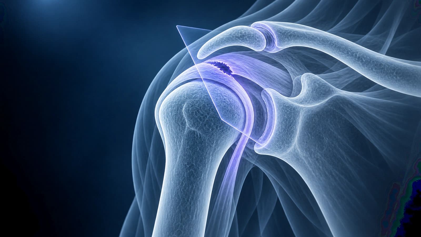
The shoulder is the most mobile joint in the body, and that mobility comes at the cost of stability. When shoulder pain strikes, MRI is the gold standard for evaluating soft tissue structures like the rotator cuff, labrum, biceps tendon, and joint capsule. Understanding what your shoulder MRI shows can help you make sense of your diagnosis and participate more actively in treatment decisions.
This guide walks you through the key structures, normal appearances, and common pathologic findings on shoulder MRI. Whether you are dealing with a rotator cuff tear, labral injury, or unexplained shoulder pain, knowing what to look for on your images can make the radiology report far less intimidating.
Shoulder MRI Views and Sequences
Shoulder MRI is acquired in three planes oriented to the shoulder joint (not the body). The coronal oblique view (aligned along the supraspinatus tendon) is the primary view for evaluating the rotator cuff. The sagittal oblique view shows the rotator cuff muscles in cross-section and is useful for assessing muscle atrophy and fatty infiltration. The axial view is essential for evaluating the labrum, glenohumeral ligaments, and subscapularis tendon.
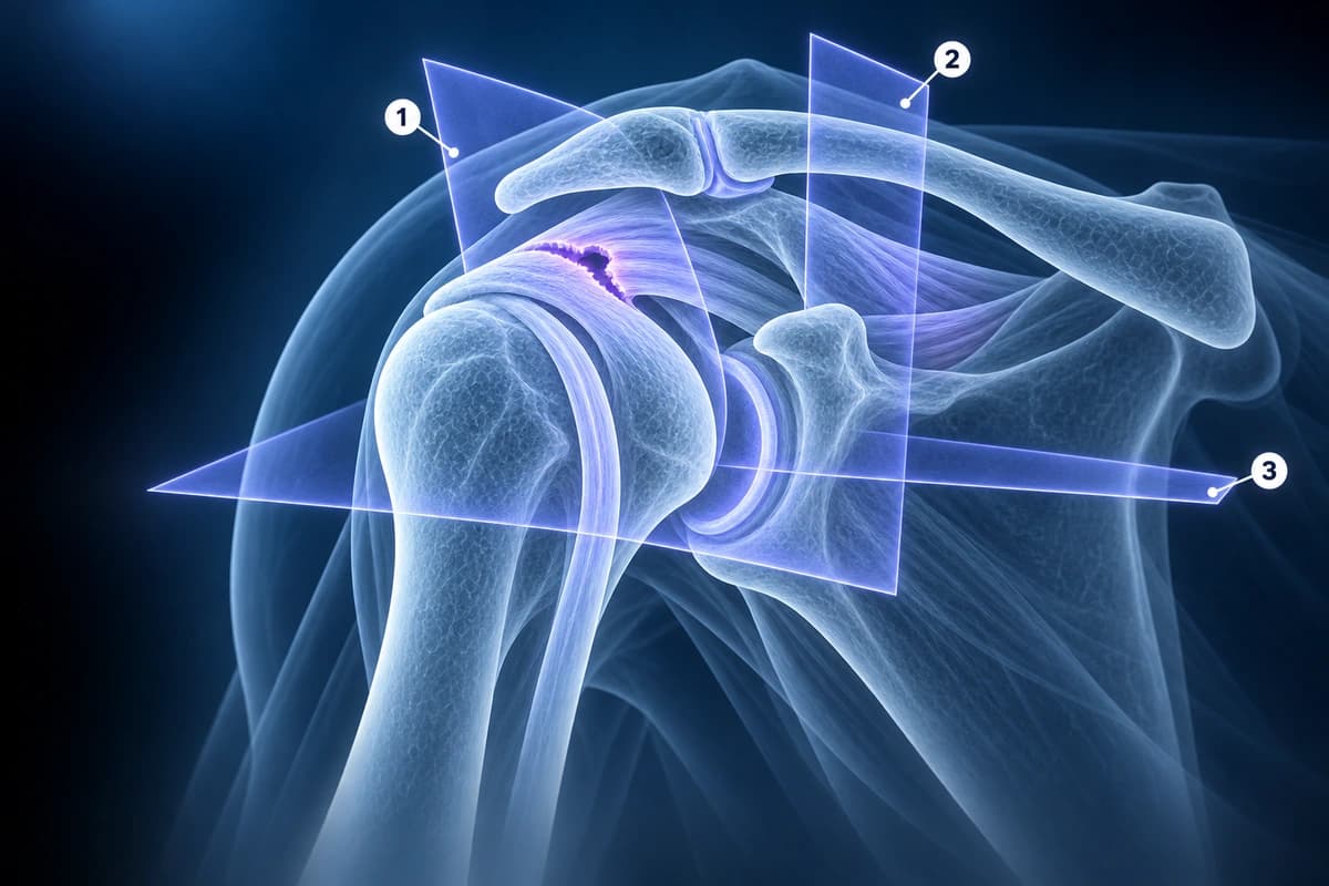
T1-weighted images show anatomy with fat appearing bright. T2-weighted fat-saturated images highlight fluid, edema, and tendon pathology as bright signal. Some protocols include MR arthrography, where contrast is injected into the joint to better delineate labral tears and partial-thickness rotator cuff tears.
The Rotator Cuff
The rotator cuff consists of four tendons: supraspinatus (most commonly torn), infraspinatus, teres minor, and subscapularis. On coronal oblique images, the supraspinatus tendon appears as a dark band extending from the muscle belly to its insertion on the greater tuberosity of the humerus. A normal tendon is uniformly dark (low signal) with a smooth, convex superior surface.
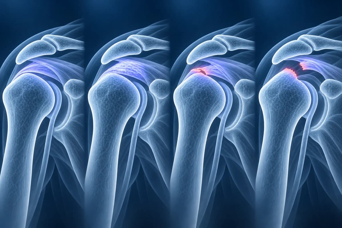
Tendinopathy appears as thickening and intermediate signal within the tendon without a discrete defect. Partial-thickness tears show focal bright signal on T2 images that does not extend through the full tendon thickness. Full-thickness tears show bright fluid signal extending completely through the tendon, often with tendon retraction. For detailed grading systems, see our article on rotator cuff tear classification.
The Labrum
The glenoid labrum is a fibrocartilaginous ring that deepens the shallow glenoid socket and provides an attachment point for the glenohumeral ligaments and biceps tendon. On axial images, the labrum appears as a dark triangular structure at the rim of the glenoid. The anterior labrum is most commonly torn (Bankart lesion), typically from shoulder dislocations.
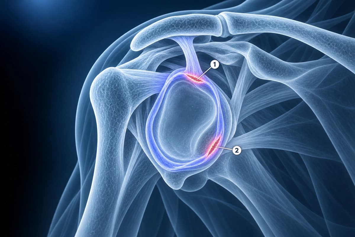
A labral tear appears as bright signal within or adjacent to the labrum, an irregular or blunted labral shape, or separation of the labrum from the glenoid rim. SLAP tears (Superior Labrum Anterior to Posterior) involve the superior labrum at the biceps anchor and are best seen on coronal oblique images. MR arthrography significantly improves labral tear detection.
Biceps Tendon, AC Joint, and Subacromial Space
The long head of the biceps tendon appears as a small, dark, round structure in the bicipital groove on axial images. Tendinopathy shows thickening and increased signal. The AC joint at the top of the shoulder commonly shows arthritis with osteophytes that can narrow the subacromial space and contribute to rotator cuff impingement. A hooked (Type III) acromion is associated with higher rates of rotator cuff tears.
Muscle Atrophy and Fatty Infiltration
When a rotator cuff tendon is torn, the associated muscle may undergo atrophy and fatty infiltration over time. These changes are best evaluated on sagittal oblique T1 images. The Goutallier classification grades fatty infiltration from 0 (no fat) to 4 (more fat than muscle). Advanced fatty infiltration (grades 3-4) is associated with poor outcomes after rotator cuff repair and may make the tear irreparable. For rehabilitation guidance, see our article on shoulder rotator cuff rehab.
Frequently Asked Questions
Can MRI detect all rotator cuff tears?
MRI detects full-thickness rotator cuff tears with 92-100% sensitivity and 85-100% specificity. Partial-thickness tears are harder to detect, with sensitivity dropping to 65-85% on standard MRI. MR arthrography improves detection of partial articular-side tears significantly.
What does tendinopathy look like on shoulder MRI?
Tendinopathy appears as tendon thickening with intermediate (gray) signal on T1 and T2 images. Unlike a tear, there is no discrete defect or bright fluid signal extending through the tendon. The tendon may appear swollen, heterogeneous, and have an irregular contour. Tendinopathy represents chronic degeneration rather than an acute tear.
Do I need an MR arthrogram or a regular MRI?
A regular MRI is sufficient for evaluating most rotator cuff tears, large labral tears, and common shoulder pathology. MR arthrography is recommended when labral tears are suspected, when evaluating partial-thickness articular-side rotator cuff tears, or in post-surgical shoulders. The decision depends on clinical presentation and what your surgeon is looking for.
What is the difference between partial and full-thickness rotator cuff tears?
A partial-thickness tear involves only part of the tendon depth — the defect does not extend completely through. A full-thickness tear extends all the way through, creating communication between the joint space and subacromial bursa. Full-thickness tears may be small (under 1 cm), medium (1-3 cm), large (3-5 cm), or massive (over 5 cm).
Sources
- Morag Y, Jacobson JA, Miller B, et al. MR imaging of rotator cuff injury: what the clinician needs to know. Radiographics. 2006;26(4):1045-1065.
- Woertler K, Waldt S. MR imaging in sports-related glenohumeral instability. Eur Radiol. 2006;16(12):2622-2636.
- Goutallier D, Postel JM, Bernageau J, et al. Fatty muscle degeneration in cuff ruptures. Pre- and postoperative evaluation by CT scan. Clin Orthop Relat Res. 1994;(304):78-83.
- Expert Panel on Musculoskeletal Imaging; Amini B, Beckmann NM, et al. ACR Appropriateness Criteria Shoulder Pain-Traumatic. J Am Coll Radiol. 2018;15(5S):S171-S188.
Related Articles
Your MRI says partial- or full-thickness rotator cuff tear? Learn what these terms, tear size and retraction mean, and how they shape treatment choices.
Evidence-based rotator cuff rehabilitation exercises, phases of recovery, and tips for optimal shoulder healing.
Your shoulder MRI mentions a SLAP tear? Learn what types I to IV mean, from labrum fraying to biceps anchor detachment, and how each type is treated.
Related Conditions
Understand your imaging before your next appointment
MRI, X-ray or CT from a CD, folder or ZIP. A PDF report or photos of the films work too.
Free to open and preview, no account needed. An optional AI report from $10 explains your images in plain language, with questions for your doctor.
Your raw files stay on this device. Only rendered images and minimal context are sent when you request an AI report.
Medical Disclaimer: This page is for informational and educational purposes only. It does not constitute medical advice, diagnosis, or treatment. AI-generated analysis may contain errors. Always consult a qualified healthcare professional for medical decisions. Full Disclaimer