
Bankart & Hill-Sachs Lesions on MRI Explained
Bankart and Hill-Sachs lesions on shoulder MRI — anterior shoulder dislocation pattern, on-track vs off-track concept, glenoid bone loss, and recurrence risk.
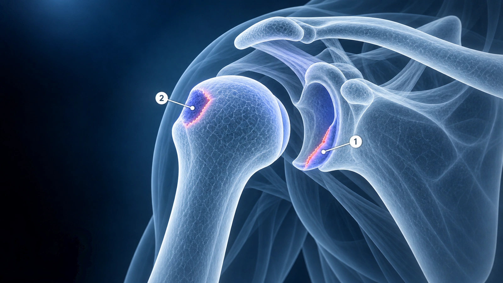
An anterior shoulder dislocation typically produces a paired bony and soft-tissue injury. The humeral head strikes the anterior glenoid rim as it dislocates, creating a compression fracture on the posterolateral humeral head — the Hill-Sachs lesion — and avulsing the anteroinferior labrum from the glenoid — the Bankart lesion. Understanding both components together is essential because their combined size and orientation determine recurrence risk, surgical strategy, and whether arthroscopic repair is sufficient or a bone-block procedure is needed.
These findings appear on almost every shoulder dislocation MRI report. This article explains what each lesion means, how radiologists measure them, and how the on-track versus off-track concept predicts whether your shoulder will dislocate again.
What Is a Bankart Lesion
The Bankart lesion is a tear of the anteroinferior labrum — the fibrocartilaginous rim that deepens the glenoid socket — caused by the humeral head levering over it during anterior dislocation. When the labrum alone is torn and pulled away from the glenoid without taking bone with it, the injury is called a soft Bankart lesion. When a fragment of the glenoid rim fractures off along with the labrum, it is a bony Bankart lesion (also called an osseous Bankart). The distinction matters because bony Bankart lesions reduce the effective articular surface of the glenoid, making the joint less stable and more likely to dislocate again — and making simple soft-tissue repair less reliable.
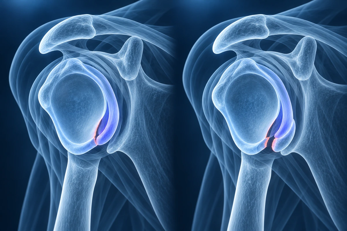
On MRI, the Bankart lesion is best visualised on axial proton-density or T2 fat-suppressed images. A soft Bankart appears as a detached or blunted anteroinferior labrum with fluid tracking between the labrum and the glenoid rim. A bony Bankart is identified by an osseous fragment medial to the anterior glenoid rim, often with surrounding marrow edema. MR arthrography — where dilute gadolinium is injected into the joint before scanning — improves detection significantly because contrast fills the labral detachment and makes small separations obvious. Conventional MRI can miss soft Bankart lesions in up to 20-30% of cases, particularly when the joint is not distended.
What Is a Hill-Sachs Lesion
The Hill-Sachs lesion is an impaction fracture of the posterolateral humeral head, created when the soft cancellous bone of the humeral head is driven against the harder glenoid rim at the moment of dislocation. The result is a groove or divot in the posterolateral humeral head that is visible on axial MRI images at the level of the coracoid process — that is the standard landmark used to identify it. On T2-weighted images, acute Hill-Sachs lesions show surrounding bone marrow edema that lights up brightly. In chronic lesions, the edema resolves and only the cortical flattening or depression remains, appearing as a step-off or groove on axial images.
The depth and width of the Hill-Sachs lesion are the two most important measurements. Deeper lesions engage the glenoid rim more easily during shoulder movements, particularly during external rotation. A shallow lesion may never cause a problem; a deep engaging lesion is a major driver of recurrent instability.
MRI Findings
The radiologist evaluates both lesions systematically. For the Bankart lesion: axial proton-density sequences are most useful; the labrum should form a smooth, dark triangle firmly attached to the glenoid rim; any separation, blunting, irregularity, or fluid undermining the labral base indicates a tear. For the Hill-Sachs lesion: axial images at the level of the coracoid show the posterolateral humeral head flattening or divot; coronal oblique images help estimate its craniocaudal height, which is used alongside width to calculate its size relative to the humeral head articular arc.
MR arthrography improves the detection of soft Bankart lesions substantially and is the preferred imaging technique when anterior instability is being worked up for surgery. Standard MRI is adequate for large bony Bankart lesions and clear Hill-Sachs defects, but misses smaller soft-tissue tears that are still clinically significant.
Glenoid Bone Loss Quantification
The amount of glenoid bone lost through repeated dislocations or a large bony Bankart fragment is one of the most important determinants of surgical planning. The glenoid is roughly pear-shaped when viewed en face. Bone loss is measured on the en-face view of the inferior glenoid — the best-fit circle is drawn over the inferior two-thirds of the glenoid, and the missing anterior arc is calculated as a percentage of that circle.
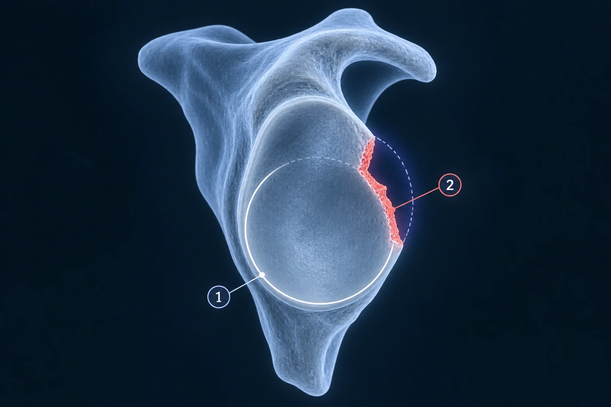
Loss exceeding approximately 20% of the glenoid surface is considered significant. At that level, the normal glenohumeral contact area is substantially reduced, and soft-tissue Bankart repair alone has unacceptably high failure rates. The PICO method (percentage of inferior circle overlap) is one established technique for this calculation on CT or MRI. CT with 3D reconstruction gives the most accurate bone-loss measurement and is often obtained in addition to MRI before surgical planning for instability.
On-Track vs Off-Track Concept
The on-track versus off-track classification combines the Hill-Sachs lesion size with the glenoid bone loss to predict whether the humeral head defect will engage the glenoid rim during normal shoulder movement — specifically during arm abduction and external rotation, the position in which anterior dislocation occurs.
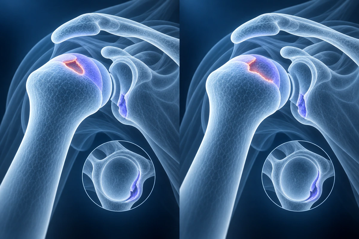
The glenoid track is the portion of the humeral head articular surface that contacts the glenoid during the full range of arm abduction-external rotation. Its width equals approximately 84% of the glenoid width minus the glenoid bone loss. If the Hill-Sachs lesion falls within this track — meaning the defect is medial enough that it never reaches the glenoid rim during normal movement — it is on-track and unlikely to cause re-dislocation. If the medial margin of the Hill-Sachs lesion extends beyond the medial border of the glenoid track — the defect reaches the rim and engages it — it is off-track. Read more about shoulder dislocation imaging.
Off-track lesions carry a substantially higher recurrence risk after soft-tissue Bankart repair than on-track lesions. Recognising an off-track bipolar lesion preoperatively changes the surgical plan.
Treatment Implications
When both the Bankart lesion and Hill-Sachs lesion are small and the Hill-Sachs is on-track, arthroscopic Bankart repair — reattaching the labrum to the glenoid with suture anchors — is the standard treatment for young, active patients after a first or second dislocation. Success rates with arthroscopic repair are excellent in this group, with recurrence rates of 5-15% in appropriately selected patients.
Significant glenoid bone loss (greater than 20%) or an off-track Hill-Sachs lesion changes the surgical approach. The Latarjet procedure — transferring the coracoid process to the anterior glenoid — augments the glenoid arc, restores the available contact surface, and provides additional stability through the sling effect of the conjoined tendon. Latarjet is preferred over soft-tissue repair in these higher-risk cases because it addresses the bone deficiency that makes soft-tissue repair fail. See also: SLAP tear classification.
For an off-track Hill-Sachs lesion without significant glenoid bone loss, remplissage is an option. In remplissage the posterior capsule and infraspinatus tendon are arthroscopically tacked into the Hill-Sachs defect, effectively filling it and preventing it from engaging the glenoid rim. Remplissage is often combined with Bankart repair and avoids the more extensive Latarjet procedure in selected patients.
Key Takeaways
- Anterior shoulder dislocation almost always produces a paired injury: Bankart lesion on the glenoid and Hill-Sachs lesion on the humeral head
- Soft Bankart = labrum only avulsed; bony Bankart = glenoid rim fragment lost — bony Bankart is more destabilising and changes surgical planning
- Hill-Sachs lesion appears as posterolateral humeral head flattening on axial MRI at the level of the coracoid
- Glenoid bone loss above 20% makes arthroscopic soft-tissue repair unreliable — Latarjet bone block is preferred
- Off-track Hill-Sachs (defect engages glenoid during arm abduction-external rotation) is a major driver of recurrent instability after Bankart repair
- Remplissage fills the Hill-Sachs defect arthroscopically and can convert an off-track lesion without large glenoid loss to a stable construct alongside Bankart repair
Frequently Asked Questions
Will my shoulder dislocate again after treatment?
Recurrence risk depends on age, activity level, and the size of the Bankart and Hill-Sachs lesions. Patients under 20 who return to contact or overhead sport after conservative treatment have recurrence rates approaching 80-90%. Arthroscopic Bankart repair reduces this substantially in appropriately selected patients (on-track Hill-Sachs lesion, minimal bone loss) — recurrence rates drop to 5-15%. When glenoid bone loss exceeds 20% or the Hill-Sachs is off-track, the Latarjet procedure achieves even lower recurrence rates of 2-8% in most series.
Is surgery necessary or can physical therapy fix these injuries?
Physical therapy cannot repair a detached Bankart lesion or reverse a Hill-Sachs impaction fracture. Soft-tissue injuries have limited intrinsic healing capacity once detached from bone. However, physical therapy is an appropriate first approach for older patients (over 40), patients with small lesions, those unwilling to undergo surgery, or those whose demands are low enough that recurrence risk is acceptable. For young active patients — especially athletes — the cumulative glenoid bone loss from repeated dislocations makes early surgical stabilisation increasingly preferred over repeated conservative management.
What exactly is the Latarjet procedure?
The Latarjet procedure transfers the coracoid process — a bony projection on the front of the scapula — together with its attached conjoined tendon to the anterior glenoid rim. It is fixed with two screws. This achieves three simultaneous effects: the bone graft extends the glenoid articular arc, making the socket effectively wider; the conjoined tendon acts as a dynamic sling against the subscapularis, blocking anterior translation in the abducted-externally rotated position; and the capsulorrhaphy performed at the same time tightens the anterior capsule. The combination makes Latarjet more resistant to failure than soft-tissue repair in patients with significant bone loss.
Does MR arthrography make a significant difference for these lesions?
Yes, particularly for soft Bankart lesions. Conventional MRI detects large bony Bankart fragments and clear Hill-Sachs divots reliably, but has only 70-80% sensitivity for soft labral detachments. MR arthrography — injecting dilute gadolinium into the joint before scanning — distends the joint and highlights even small labral separations as bright contrast signal. Sensitivity improves to approximately 90% for labral tears. If surgery is being planned for instability, MR arthrography is generally preferred over standard MRI to avoid underestimating the Bankart lesion size.
Can Bankart and Hill-Sachs lesions heal without surgery?
The Hill-Sachs impaction fracture fills in with fibrous tissue over time but the cortical depression does not fully remodel — its depth and engagement potential remain. The Bankart labral detachment does not re-attach to bone without surgical repair; the labrum may scar in a medialised position, which leaves the anterior capsule lax and the shoulder prone to repeat dislocation. For this reason, in young active patients the standard approach has shifted toward early surgical stabilisation after a first-time dislocation, rather than waiting for multiple recurrences and accumulating progressive bone loss before intervening.
Related Articles
Understand SLAP tear types I-IV on shoulder MRI, including superior labrum fraying, biceps anchor detachment, bucket-handle tears, biceps tendon extension, and treatment implications.
Understand your shoulder MRI report, key anatomical structures, and what common findings mean.
Related Conditions
Ready to analyze your imaging? Upload your MRI or X-ray for AI-powered analysis
Upload your MRI or X-ray DICOM files for private, AI-powered analysis. 4 models analyze independently — raw scan files stay in your browser.
Start AnalysisMedical Disclaimer: This page is for informational and educational purposes only. It does not constitute medical advice, diagnosis, or treatment. AI-generated analysis may contain errors. Always consult a qualified healthcare professional for medical decisions. Full Disclaimer