
Supraspinatus Tendinosis on MRI: Tendinopathy vs Tear
Your shoulder MRI says supraspinatus tendinosis or tendinopathy? Learn how it differs from a partial- or full-thickness tear and what to ask your doctor.
“Supraspinatus tendinosis” in an MRI report
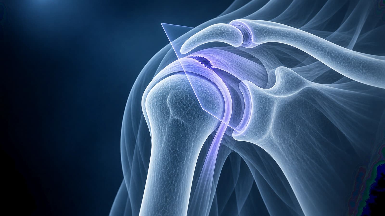
Supraspinatus tendinosis, also written tendinopathy or tendinitis, means the tendon on top of the shoulder looks thickened or shows intermediate signal on MRI without a gap through its fibers. Tissue studies show this signal reflects degeneration rather than active inflammation. A tear, by contrast, shows fluid-bright signal that reaches a tendon surface.
How the finding is worded
- “Mild supraspinatus tendinosis without tear”
- “Supraspinatus tendinopathy with intrasubstance signal”
- “Insertional tendinosis with a small partial-thickness articular-sided tear”
- “Tendinosis versus low-grade partial-thickness tear”
Tendinosis or tear: how the images differ
Radiologists look at how bright the signal is and where it goes. Intermediate signal that stays inside a thickened tendon fits tendinosis. Signal as bright as fluid that reaches the joint-side or bursal-side surface fits a partial-thickness tear, and signal crossing the whole tendon fits a full-thickness tear. Some patterns overlap, which is why a report may say “tendinosis versus low-grade partial tear”.
If a tear is reported, its depth and size are graded as explained in partial vs full-thickness rotator cuff tears.
Is tendinosis serious?
Rotator cuff changes become common with age, even in shoulders that do not hurt: in one MRI study of people without symptoms, 54% of those over 60 had a cuff tear. So tendinosis on a report is interpreted alongside your pain, strength and examination, and it often sits next to findings such as subacromial narrowing or bursitis.
What to ask at your follow-up
- Is this tendinosis only, or is there also a partial tear?
- How much of the tendon is involved, and is it at the insertion on the bone?
- Does the scan also show bursitis or calcium deposits?
- Which exercises or activity changes fit this finding?
Sources
- Kjellin I, Ho CP, Cervilla V, et al. Alterations in the supraspinatus tendon at MR imaging: correlation with histopathologic findings in cadavers. Radiology. 1991;181(3):837-841.
- Rafii M, Firooznia H, Sherman O, et al. Rotator cuff lesions: signal patterns at MR imaging. Radiology. 1990;177(3):817-823.
- Morag Y, Jacobson JA, Miller B, et al. MR imaging of rotator cuff injury: what the clinician needs to know. Radiographics. 2006;26(4):1045-1065.
- Sher JS, Uribe JW, Posada A, et al. Abnormal findings on magnetic resonance images of asymptomatic shoulders. J Bone Joint Surg Am. 1995;77(1):10-15.
Related Articles
Your MRI says partial- or full-thickness rotator cuff tear? Learn what these terms, tear size and retraction mean, and how they shape treatment choices.
A patient's guide to the shoulder MRI report: rotator cuff, labrum, biceps and bursa terms in plain words, and what common findings usually mean.
Related Conditions
Understand your imaging before your next appointment
MRI, X-ray or CT from a CD, folder or ZIP. A PDF report or photos of the films work too.
Free to open and preview, no account needed. An optional AI report from $10 explains your images in plain language, with questions for your doctor.
Your raw files stay on this device. Only rendered images and minimal context are sent when you request an AI report.
Medical Disclaimer: This page is for informational and educational purposes only. It does not constitute medical advice, diagnosis, or treatment. AI-generated analysis may contain errors. Always consult a qualified healthcare professional for medical decisions. Full Disclaimer